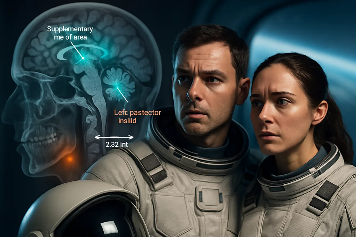
Imagine returning from a year in orbit only to find that your brain has moved a few millimeters within your skull. The striking MRI evidence of brain shifts after spaceflight demands attention, because those millimeters correspond to altered motor control, balance deficits, and a recovery timeline that current strategies may underestimate.
Why brain shifts after spaceflight matter for mission safety
This new MRI study of 26 astronauts shows consistent upward and backward displacement of the brain, with some regions moving unevenly by several millimeters. Those microshifts are not trivial: the supplementary motor area showed an average upward displacement of 2.52 millimeters after year-long missions, and shifts in the posterior insula correlated with measurable balance decline.
Consequently, brain displacement is not merely an academic curiosity. It intersects directly with operational performance, rehabilitation planning, and the risk calculus for longer lunar or Mars expeditions. If sensory integration and motor planning centers relocate relative to the skull, astronauts may face persistent deficits even as muscles and bones recover.
What the MRI evidence actually shows about intracranial fluid shifts
The researchers anchored brain measurements to the skull, separating global upward drift from regional stretching and compression. This approach reveals nonlinear deformation rather than a simple rigid shift. Some cortical areas compress, others stretch, and left-right asymmetries blur in whole-brain averages.
Moreover, the study compared astronauts with a 60-day head-down tilt bed-rest analog to probe which features of space exposure are reproducible on Earth. The analog reproduced parts of the pattern, especially backward displacement, but failed to match the magnitude and directional balance of real microgravity, particularly the stronger upward component observed after true spaceflight.
Regional hotspots and functional stakes
Topical regions near the vertex and sensorimotor strips showed the largest movements, and the posterior insula emerged as a functionally relevant hotspot. Larger posterior insula displacement predicted worse performance on the Sensory Organization Test, linking anatomy to balance and postural control deficits.
Thus, MRI-detected positional changes are not only structural markers. They carry measurable consequences for how crewmembers sense their head in space, coordinate locomotion, and stabilize themselves after landing. Actions that rely on fine timing and integration of vestibular and proprioceptive cues become vulnerable.
Argument: Bed-rest analogs are useful but insufficient as a policy foundation
Analog studies like AGBRESA are indispensable for controlled experimentation and countermeasure testing, yet the evidence argues they cannot be treated as full substitutes for spaceflight. The bed-rest protocol reproduced some aspects of the deformation and recovery curves but did not replicate the full upward displacement pattern seen in orbital missions.
Therefore, agencies that base mission planning primarily on analog results risk underpreparing crews for direction-specific brain adaptation. Analog-derived timelines could underestimate how long vestibular and sensorimotor systems remain altered after a long mission, and they may misjudge which countermeasures actually mitigate crucial regional shifts.
Why this distinction matters for countermeasure design
For example, artificial gravity as applied in the AGBRESA campaign did not produce statistically significant protection at the tested doses. That result does not close the door on artificial gravity, but it does critique simplistic implementations. If brain deformation is nonlinear and region-specific, a one-size-fits-all short-radius centrifuge regimen may not address the hotspots most relevant to balance and motor planning.
Consequently, countermeasure development should couple realistic spaceflight MRI phenotypes with mechanistic testing in both analogs and flight, rather than extrapolating from short-duration or low-dose analog interventions alone.
Rehabilitation and recovery: timelines that must be recalibrated
The postflight scans in the prospective astronaut subgroup show partial but uneven recovery over six months. Vertical displacement largely rebounded by six months, yet backward displacement and some regional deformations persisted. These recovery dynamics are not trivial for mission-readiness tasks upon return.
In operational terms, balance testing, gait retraining, and daily functional tasks performed in the weeks after landing occur while the nervous system is still reorganizing. Rehabilitation protocols that assume rapid neural reset may be optimistic, and risk management must include longer-term neurorehabilitation for crewmembers returning from long-duration missions.
Practical implications for astronaut care
First, preflight baseline imaging and serial postflight MRI need to be routine to map individual deformation and recovery trajectories. Second, vestibular and sensorimotor training should be prioritized before, during, and after flight with protocols designed to address region-specific vulnerabilities, not just gross deconditioning.
Finally, rehabilitation teams should integrate neuroimaging data into functional assessments. If posterior insula displacement predicts balance decline, vestibular therapy can be targeted and progress objectively tracked using both performance metrics and anatomical markers.
Policy and research priorities that follow from the argument
Given the evidence, space agencies and research funders should adopt three interlocking priorities. First, increase the sample size and diversity of longitudinal astronaut neuroimaging, including more one-year missions and multiple postflight timepoints. More data will allow separation of individual variability from systematic effects.
Second, invest in higher-fidelity analogs and in-flight countermeasure trials that are tailored to the observed deformation profiles. This could mean longer-duration centrifuge exposure, different gravity gradients, or integrated vestibular stimulation protocols tested in both analog and flight contexts.
Third, align operational timelines with neurorecovery realities. Mission planners should build guardrails for postlanding activities, stagger return-to-duty assessments, and fund sustained rehabilitation contingencies for crews who show persistent deformation in critical regions.
Translating research into equipment and training
Practical translation includes redesigning spacecraft exercise regimens to incorporate targeted vestibular challenges, developing in-flight diagnostic tools for intracranial fluid monitoring, and optimizing suit or seat designs to mitigate fluid shifts during reentry. These steps are not extravagant; they are logical responses to millimeter-scale brain displacement that influences function.
Moreover, mission medical teams should be prepared to personalize countermeasures. Individual variation in deformation and recovery suggests that flexible protocols will succeed where uniform prescriptions fail.
Balancing optimism and caution as missions grow longer
On the one hand, the brain demonstrates plasticity and shows substantial recovery in many dimensions. On the other hand, the persistence of some deformations and the clear link to balance deficits counsel caution. Ambitious plans for lunar outposts or Martian transits must integrate neurobiological risks with the same rigor applied to radiation, muscle, and bone health.
Therefore, treating brain displacement as a first-class operational risk is not alarmist. It is a rational alignment of medical planning with observable anatomical and functional consequences of living in microgravity for extended periods.
Actionable steps for researchers and mission planners
Prioritize longitudinal MRI protocols with skull-anchored measures to detect regional deformation early. Complement imaging with vestibular and gait assessments that are sensitive to posterior insula and supplementary motor area dysfunction. Test countermeasures at doses and durations likely to affect regional fluid dynamics, not just global metrics.
Furthermore, design rehabilitation pathways that assume weeks to months of ongoing neural recovery, and fund adaptive training devices that can be tailored to each crewmember’s imaging profile. These are feasible, high-impact investments compared with the human costs of impaired balance or delayed operational readiness after long missions.
Over decades of human spaceflight, millimeters inside the skull will accumulate into mission outcomes. The emerging evidence about brain shifts after spaceflight argues forcefully for integrated research, tailored countermeasures, and rehabilitation plans that treat regional neuroanatomy as central to crew health and performance. By aligning imaging, analog testing, and operational timelines, agencies can protect both the bodies and the brains of future explorers, turning a small anatomical shift into a manageable risk rather than an operational surprise.

Dr. Morgan directed the Archives Program from 2014 to 2017, gaining extensive experience in research documentation, information management, and the preservation of scholarly resources. Throughout her career, she has worked closely with academic publications and research materials, developing expertise in evaluating scientific sources and communicating complex topics to broad audiences.
Her primary areas of specialization include scientific publishing, research communication, editorial review, and the translation of technical research into accessible educational content. She has contributed to projects involving space science, astronomy, environmental science, history, archaeology, and emerging scientific discoveries, always emphasizing accuracy, transparency, and the responsible presentation of evidence.
As Editorial Director of Muskurahat.us, Dr. Morgan leads the editorial review process for scientific articles, ensuring that content is based on reputable sources, peer-reviewed research whenever available, and publications from recognized universities, research institutions, and international scientific organizations.
She is committed to promoting scientific literacy through clear, engaging, and well-documented articles that help readers better understand scientific discoveries and their impact on society. Her editorial philosophy is founded on accuracy, intellectual integrity, independent journalism, and continuous learning as scientific knowledge evolves.
Through her work at Muskurahat.us, Dr. Morgan supports the publication of trustworthy scientific content that makes complex research accessible to readers around the world while maintaining rigorous editorial standards.